
How to Use a Microscope (Properly) Step by Step New York Microscope
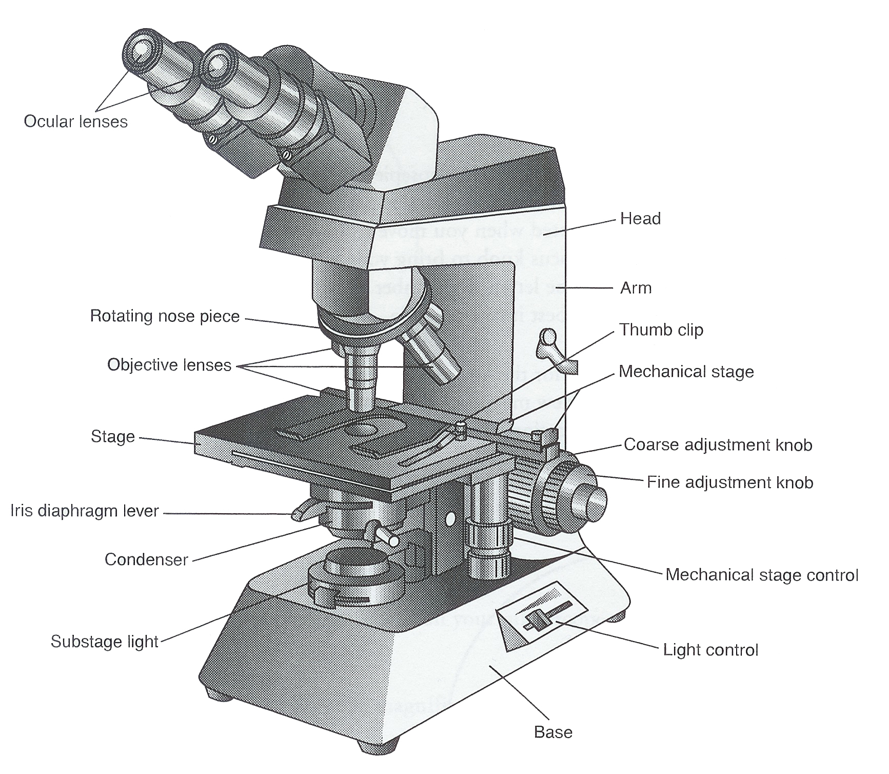
Iris diaphragm: Adjusts the amount of light that reaches the specimen. Condenser: Gathers and focuses light from the illuminator onto the specimen being viewed. Base: The base supports the microscope and it's where illuminator is located. How Does a Compound Microscope Work?
Cells and Microscopes
Record the microscope images using labelled diagrams or produce digital images. When first examining cells or tissues with low power, draw an image at this stage, even if going on to examine the.

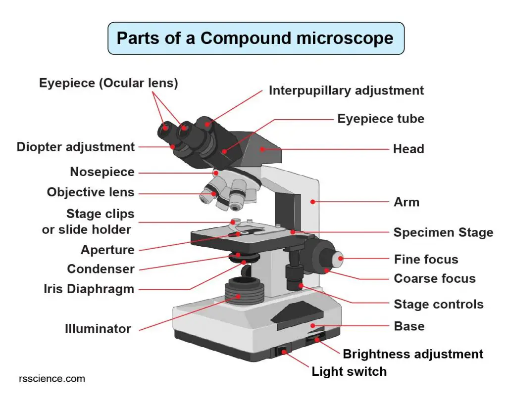
Parts of a Compound Microscope Labeled (with diagrams) Medical
The web page titled "Parts of a Microscope with Labeled Diagram and Functions" has the following key takeaways: 🔍 The microscope is an essential tool for scientists, researchers, and medical professionals. 🧬 The main function of a microscope is to provide a magnified view of small objects or organisms, such as bacteria, cells, or.
Simple Microscope Definition, Principle, Parts, And Uses » Microscope Club
A Study of the Microscope and its Functions With a Labeled Diagram - Science Struck A Study of the Microscope and its Functions With a Labeled Diagram To better understand the structure and function of a microscope, we need to take a look at the labeled microscope diagrams of the compound and electron microscope.
Clipart microscope parts labeled WikiClipArt
The optical microscope often referred to as the light microscope, is a type of microscope that uses visible light and a system of lenses to magnify images of small subjects. There are two basic types of optical microscopes: Simple microscopes. Compound microscopes. The term "compound" in compound microscopes refers to the microscope having.

Parts of a Microscope The Comprehensive Guide Microscope and
Parts of the Microscope (Labeled Diagrams) By Editorial Board December 14, 2022 The microscope is one of the must-have laboratory tools because of its ability to observe minute objects, usually living organisms that cannot be seen by the naked eyes. It is categorized into two: simple and compound microscopes.

Parts of a microscope with functions and labeled diagram
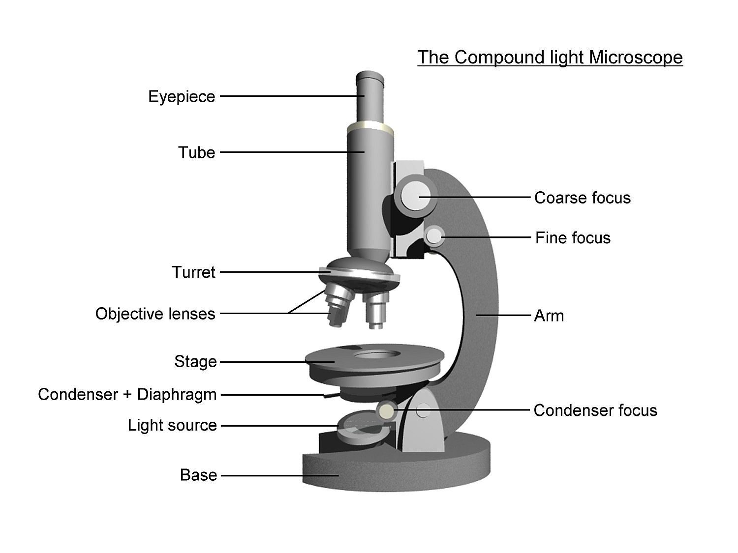
Figure: Diagram of parts of a microscope. There are three structural parts of the microscope i.e. head, arm, and base. Head - The head is a cylindrical metallic tube that holds the eyepiece lens at one end and connects to the nose piece at other end. It is also called a body tube or eyepiece tube.
Microscope Labelled Diagram Gcse Micropedia Gambaran
Compound Microscope Parts - Labeled Diagram and their Functions Microscopes / By Rachael Sharing is caring! This article will review the structure of a compound microscope and explain to you how each part works to give us the magnification images. This article covers An overview of microscopes What is a "compound microscope"?
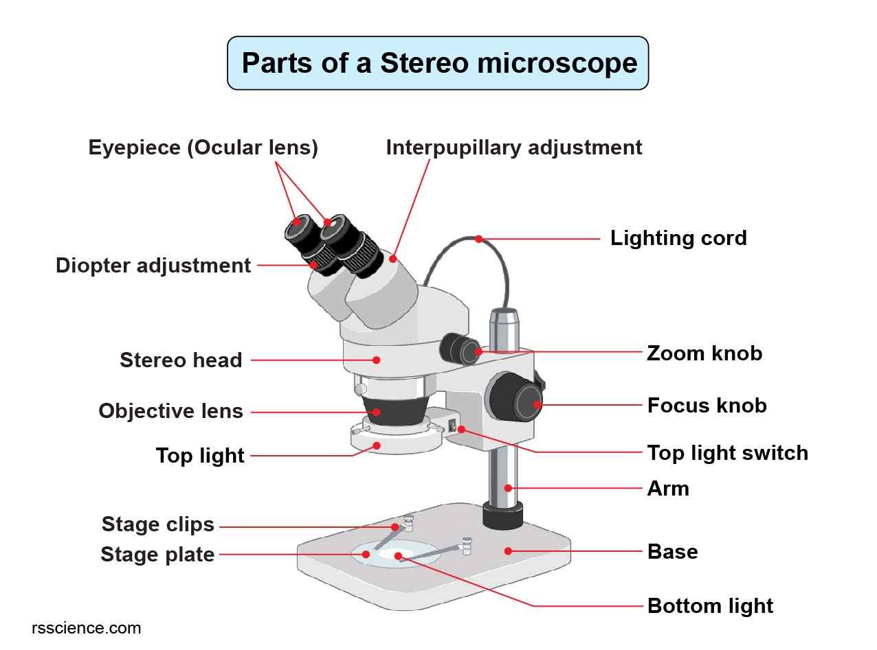
Parts of Stereo Microscope (Dissecting microscope) labeled diagram
A microscope is an instrument that magnifies objects otherwise too small to be seen, producing an image in which the object appears larger. Most photographs of cells are taken using a microscope, and these pictures can also be called micrographs. From the definition above, it might sound like a microscope is just a kind of magnifying glass.
Compound Microscope Parts Labeled Diagram and their Functions (2023)
Meiji MT-30 Binocular Microscope - Rechargeable. $618.55. Labomed 9135010 CxL Binocular Cordless Microscope, 4x, 10x, 40x Objectives, LED Illumination. $741.00. ACCU-SCOPE EXM-150-MS Monocular Cordless Microscope with Mechanical Stage, Rechargeable. $351.90. Get relevant offers, the latest promotions, and articles from New York Microscope.
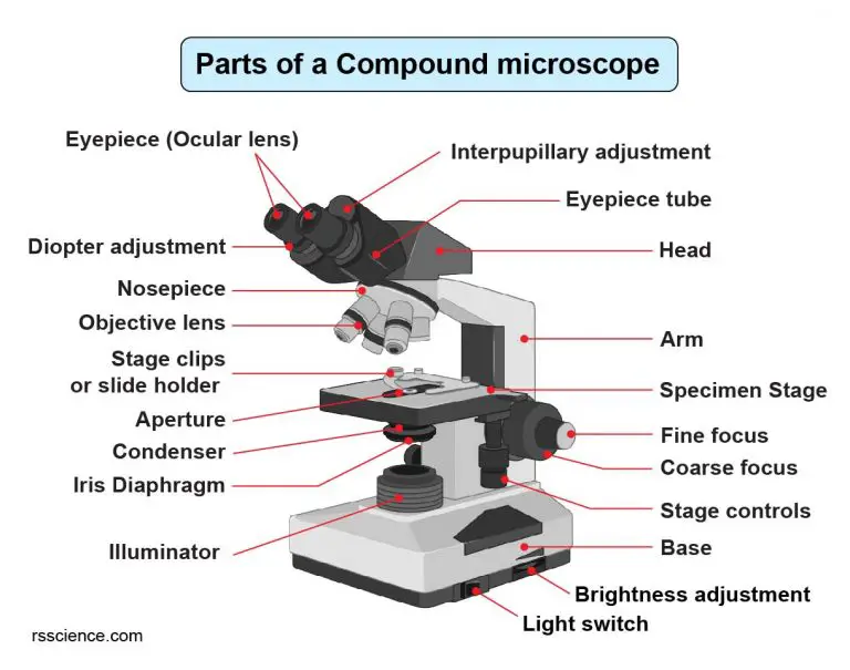
Compound Microscope Parts Labeled Diagram and their Functions Rs
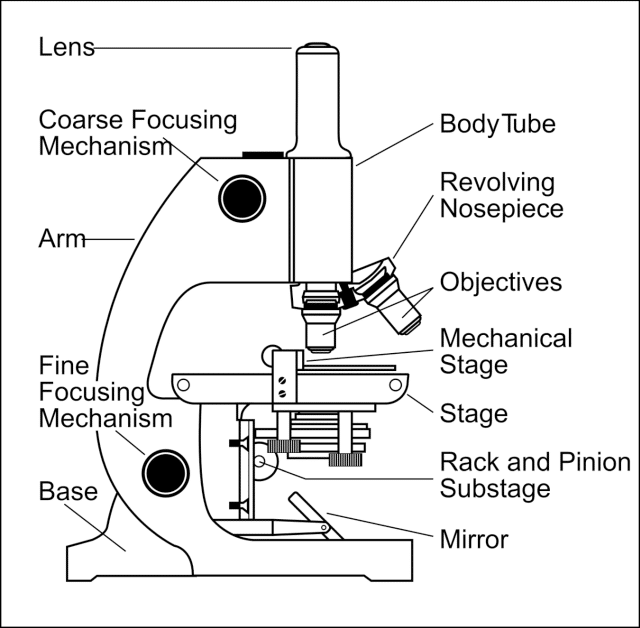
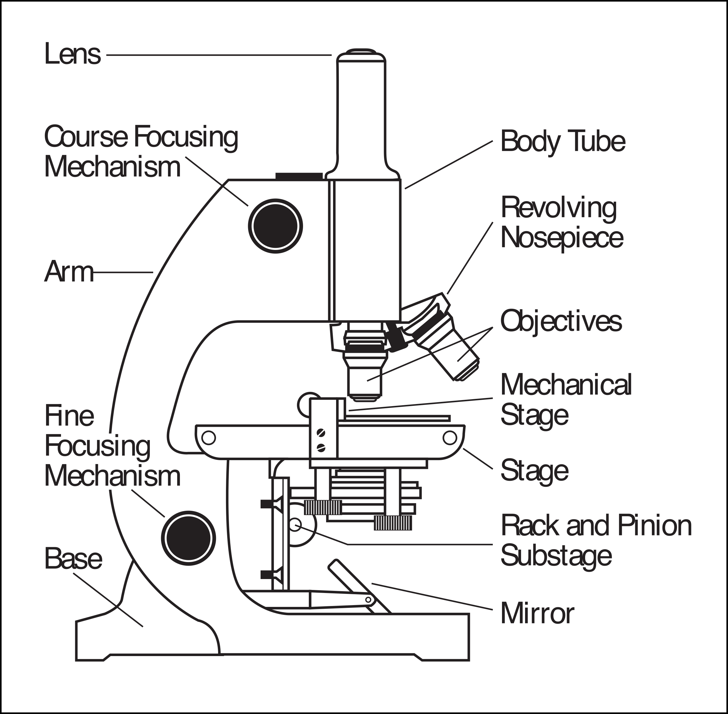
Labeling the Parts of the Microscope This activity has been designed for use in homes and schools. Each microscope layout (both blank and the version with answers) are available as PDF downloads. You can view a more in-depth review of each part of the microscope here. Download the Label the Parts of the Microscope PDF printable version here.
1.5 Microscopy Biology LibreTexts
A microscope is a piece of laboratory optical equipment used to magnify small things that are too small for the details to be seen by the naked eye. The microscope is the microbiologist's most basic tool, and every microbiology student needs some background knowledge on parts of a microscope and how microscopes work.
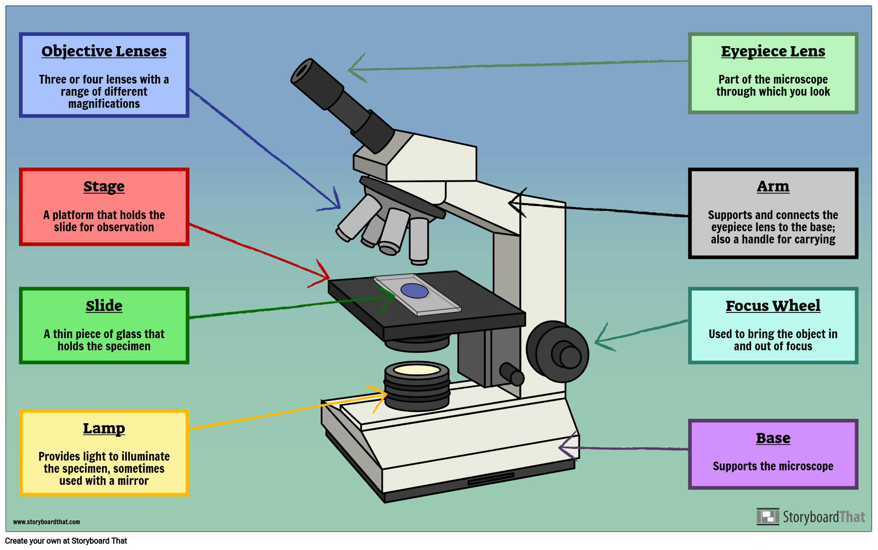
Labelled Microscope with Functions Storyboard by oliversmith
Create a poster that labels the parts of a microscope and includes descriptions of what each part does. Click "Start Assignment". Use a landscape poster layout (large or small). Search for a diagram of a microscope. Using arrows and textables label each part of the microscope and describe its function. More options.
Parts Parts And Functions Of A Microscope
ACTIVITY Microscope parts In this activity, students identify and label the main parts of a microscope and describe their function. By the end of this activity, students should be able to:. READ MORE MORE Use this interactive to identify and label the main parts of a microscope. Drag and drop the text labels onto the microscope diagram.
How to Use a Microscope
Light and electron microscopes allow us to see inside cells. Plant, animal and bacterial cells have smaller components each with a specific function. We need microscopes to study most cells.

Microscope diagram Tom Butler Technical Drawing and Illustration
Simple Microscope Diagram (Parts) with Labels Frequently Asked Questions Definition and Working principle Simple microscope is a magnification apparatus that uses a combination of double convex lens to form an enlarged, erect image of a specimen.
